SS#_________________________
Grade_________/100
MatSE 430
1st Exam - 1996
Par time - 21.5 min.
2.5 min.
2. (25) (not covered this year)For an orthorhombic material with a =6.54, b= 7.23, c = 9.18 Å, and n(alpha) = 1.38, n(beta) = 1.41, and n(gamma) = 1.52, determine the permitted vibration directions and associated refractive indices for the incident light normal to the (210) crystal face. Show all of your calculations. Show the results (i.e., the beam direction, the permitted vibration directions and associated refractive indices, and all the angles necessary to define these directions) in a perspective drawing below.
8 min.
3. (15) The micrograph below (not shown here) of a certain material was taken at 400X using a reflected light microscope and white light. Describe what is seen in this microstructure.
Now, obviously, the structure is not very clear, even though the microscope was correctly focused. What changes could be made with respect to the setup of the microscope to possibly obtain a clearer image? Explain why, using the appropriate equation.
2.5 min.
4a. (20) Describe how characteristic x-rays are produced in an x-ray tube. What control the intensity of the x-ray beam? The wavelength?
Use a drawing to describe what goes on in the atoms of the anode material.
4b. How does the process for the production of fluorescent x-ray in a diffractometer specimen differ from the above?
2.5 min.
5. (25) Index, by any appropriate means, these diffraction data (interplanar spacings) for a cubic materials and give the Bravais lattice and a representative lattice parameter. Show all calculations.
6 min.
Bravais lattice__________________
a = ______________
3.184 Å
2.603
2.252
2.015
1.835
1.500
1.421
SS#_________________________
Grade_________/100
MatSE 430
2nd Exam - 1996
Par time - 21 min.
(b) What is one major advantage of an electromagnetic lens for electrons, versus an optical lens for light (photons)?
(c) What is one major disadvantage of an electromagnetic lens for electrons, versus an optical lens for light (photons)?
2 min.
2. (15) For each of the materials problems listed below, indicate which of the various electron microscopy techniques would be most useful. In each case, briefly explain why.
· scanning electron microscopy - with a secondary electron detector (SEM-SE)
· scanning electron microscopy - with a backscattered electron detector (SEM-BS)
· transmission electron microscopy (TEM)
(a) examining the regularity of the surface pits on a CD that digitally encode the audio information (prior to the addition of any protective surface layers)
(b) characterizing the density of crystallographic defects (dislocations) in a rolled steel
(c) determining grain size on the polished and then etched surface of a ceramic material
(d) nondestructive analysis to determine whether only a single phase, or multiple phases, are present in an annealed knife blade
2.5 min.
3. (15) Electron detectors used in SEM often have a metal mesh in front of a scintillator. Is this mesh biased positively, negatively, or not biased for: (Briefly explain why in each case.)
(a) secondary electron detection?
(b) backscattered electron detection?
1.5 min.
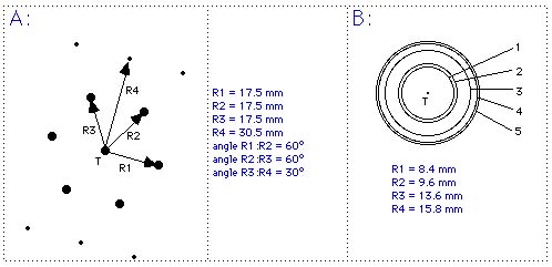
4. (15) Of what phase(s) do you suspect this sample is composed? List file d's and I's to the right of the data in a separate column for each phase. See sheet near back of exam (not shown here).
4.705 10
4.262 18
3.988 18
3.915 43
3.875 100
3.815 52
3.785 32
3.710 21
3.472 78
3.427 42
3.133 46
3.120 15
2.907 30
2.788 25
2.594 57
2.332 25
4 min.
5. (25) (not covered this year) ZrSi2 is Cmcm, a = 3.721, b = 14.68, c = 3.683 Å with Zr atoms in 4c (y = 0.102) and Si atoms in 4c (y = 0.752 and 0.446). Calculate I(111) CuKalpha radiation. Ignore scale factor, absorption, and temperature factor. LP = (1 + cos2 2theta)/(sin2 theta cos theta). (Give correctly the summation signs and summation limits! Carefully show all calculations and fill in the table. Interpolate for 's.)
(hkl) dhkl (sin theta hkl)/lambda F(hkl)
Zr:
(sin theta)/lambda 0.0 0.1 0.2 0.3 0.4 0.5 0.6 0.7
40 36.3 30.8 26.0 22.1 19.7 17.5 15.6
Si:
(sin theta)/lambda 0.0 0.1 0.2 0.3 0.4 0.5 0.6 0.7
14 11.35 9.4 8.2 7.15 6.1 5.1 4.2
8 min.
6. (15) (a) Circle those items listed below which are not included in the data in the PDF.
unit cell dimensions
optical constants
space group
atom positions
density
2theta values
melting points
mass absorption coefficients
(b) Discuss briefly how the ICDD obtains the powder diffraction patterns for the PDF.
(c) (not covered this year) What is the meaning of the factor e-2M? How does it influence the intensities of reflections and why (use a drawing which compares ideal case to that in which this factor applies to discuss why)?
Grade_________/100
Mat Sc 430
Final Exam - 1996
Par time - 33.75 min.
Pb2O: simple cubic, a = 5.38 Å
TiO: face-centered cubic, a = 4.18 Å
Pb~2(ZrxTi2-x)O~7 (pyrochlore): face-centered cubic, a = 10.40 Å
Pb(ZrxTi1-x)O3 (perovskite): simple cubic, a = 4.04 Å 18 min.
In the area shown, there are two distinct kinds of regions: the dark roughly circular patches, and the lighter material in between. The diffraction patterns shown on the next page (simulated, but similar to the real ones, which don't reproduce well; transmitted beam indicated by "T") are taken at an accelerating voltage of 200 kV and a camera length of L=2000 mm from one of the dark circular areas (A) and from a light area between these circular areas (B). Answer the following questions based on these data. Show your work and/or explain your reasoning in each case. (In analyzing the diffraction patterns, use the marked values of the measurements even if the actual spacings and angles come out a little differently on your exam sheet.)
Here are some equations that you may or may not find useful:
 (a) From the diffraction pattern (A), determine whether the dark circular
area is the desired perovskite phase, one of the other three phases listed,
or something else.
(a) From the diffraction pattern (A), determine whether the dark circular
area is the desired perovskite phase, one of the other three phases listed,
or something else.
(b) From the diffraction pattern (B), determine whether the material in the lighter regions is the desired perovskite phase, one of the other three phases listed, or something else.
(c) Determine the orientation of the phase in the dark circles by fully indexing diffraction pattern A and then finding the zone axis direction. (This crystallographic direction must then be perpendicular to the film surface, at least for this grain, since it is parallel to the electron beam direction!)
(d) Is the contrast between the two major kinds of areas most likely a result of absorption (scattering) contrast, diffraction contrast, or phase contrast? Explain why.
2. (10) Name the three fundamental components of a thermionic electron gun and the three primary parameters that are varied (gun controls). For the latter, indicate exactly what is varied in each case and how it relates to the three basic components. What is meant by "saturation" with respect to the electron gun? Which of the three gun controls is adjusted in order to reach saturation? 2 min.
3. (8) Circle, check, or otherwise indicate (clearly!) the most correct completions of the following statements about scanning electron microscopy. IMPORTANT NOTE: to discourage random guessing, incorrect answers will REDUCE your score on the exam by 0.5 points each! 1.5 min.
(a) Increasing the working distance will:
- improve image resolution
- decrease the image signal-to-noise ratio
- increase astigmatism of the electron beam
- increase the maximum field of view and the depth of field
(b) Increasing magnification:
- reduces sample damage
- increases the area scanned on the sample
- reduces the area scanned on the sample
- improves image resolution
(c) The objective lens controls the:
- focus of the image
- electron beam current
- image brightness
- depth of field of the image
(d) Reducing the size of the final aperture will:
- improve resolution
- increase the total electron current on the specimen
- increase the depth of field
- reduce the area scanned on the sample
4. (15) (not covered this year) A material with lattice constants a = 5.623, c = 14.103 Å, and a = b = 90°, g = 120°, has n(alpha) = 1.42, n(beta) = 1.65, and n(gamma) = 1.85. Determine the permitted vibration directions and associated refractive indices for the incident light normal to the (011) crystal face of a single crystal of the material.
1. Carefully determine the angles which define the direction of the light beam using diagrams.
2. Construct the perspective diagram on the next page which shows the beam direction, the permitted vibrational directions and associated refractive indices, and all the angles necessary to define these directions.
3. Finally, determine the two unknown n's and mark them on the diagram.
Show all of your calculations. The 3-D drawing must be shown. 6 min.
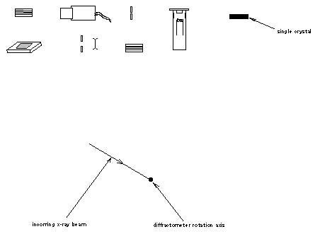
5. (15) Using all of the components given below, construct a conventional powder diffractometer by drawing them in their precise relative locations and orientations. When your drawing is complete, carefully show the focusing circle. 1 min.
6. (15) The IR spectrum given below (not shown here) is for a polymer. See if you can identify the polymer. Since you may not be very familiar with polymers, you may give your answer as any of the following: a) the name of the polymer, b) a sketch of what you think the structure of the polymer molecule may look like, and/or c) a discussion of the components of the polymer molecule and how they are put together. Explain your choice. 1.25 min.
7. (5) In x-ray, electron, and neutron diffraction studies, a generalized form of the structure factor, F(h), turns out to be the Fourier transform of the electron density, r(X). Write the equation which expresses this relationship. Use the exponential form of the kernel. 0.75 min.
8. (12) What do you consider to be the major consideration (or problem) in specimen preparation (solids), and why, for : 3.25 min.
reflected light microscopy
x-ray diffraction
IR spectroscopy