SS#________________________________
Grade____________________/10
MAT SC 430 - HOMEWORK # 6 - Due 10/5/00
Optical microscopy
1. Visit the Olympus BHM microscope in 202 Steidle (NW corner, main room). Make a diagram of the optics of this microscope (don't make a drawing of the instrument) as viewed from the right side and identify all of the components. What magnifications and NAs are possible with this instrument? Immediately above the power connection to the lamp is a knob which, when turned does something. What? For what purpose? Locate the rotatable adjustment marked F. What does it do? For what purpose? Finally, what is the purpose of using the slide adjustment above the lens turret? (Uppermost knob, slides horizontally right and left.)
You should find a specimen marked MatSC 430 nearby to view. Warning!!! Do not let the bottom lens touch the specimen. The best procedure for focusing is to adjust the microscope until the lens is seen to be just above the specimen surface. Then look into the eyepieces and adjust by raising the bottom lens.
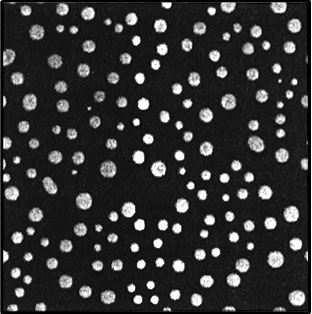
2. For the microstructure shown below, calculate the volume fraction of the white phase by a) point counting and b) through the use of the program NIH Image. For point counting, use two grid sizes; give grid sizes, calculations, and results for both measurements. For the computer calculation, use the Mac in 202 Steidle or one of those in 106 Steidle. The filename for the image is "430 image". Open Image, open "430 image", density slice, adjust LUT, make binary, analyze particles, show results, measure. You will have to add the areas of over 100 particles and then divide by the total area; you may wish to store the results in a file to import into Excel or word for these calculations. Please discard all such files when you are finished. Briefly compare results from the point counting and image analysis techniques. Consult About NIH Image for help.